Unlocking the Structure and Function of Cytochrome P450s
January 13, 2025
By Toni Shears 
Of the 57 human cytochrome P450 enzymes, about half control normal physiological processes such as blood pressure, reproduction, inflammation and more. They produce steroid hormones and bile acids, fatty acids, and vitamins. When these enzymes don’t do enough — or when they do their jobs too well — it often leads to disease. That makes cytochrome P450s prime targets for drug development. Often the goal is to design a drug that fits into the active site cavity of a P450 enzyme, like a key sliding into a lock, to either inhibit or boost P450 function.
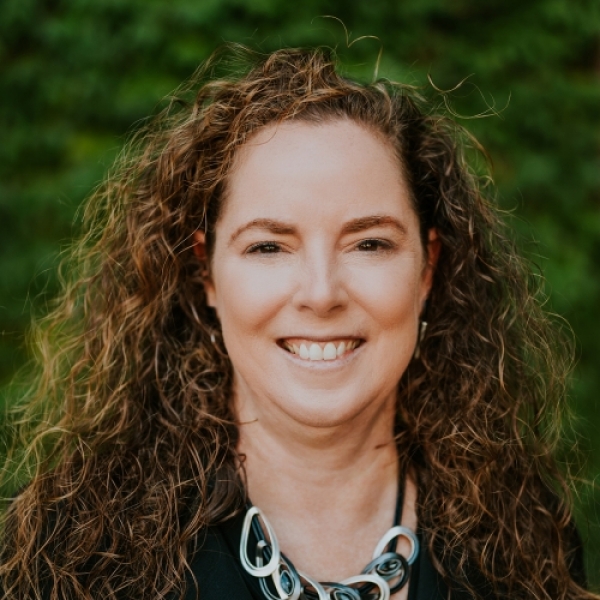
Professor Emily Scott’s drug discovery research painstakingly maps the structure and function of human cytochrome P450s. Substantial effort goes into defining the shape, size, and chemical characteristics of a P450 active site (the lock) to tell chemists how to compose the drug (key) to boost or suppress P450 function.
Her lab makes human P450 proteins in bacteria, isolates and crystallizes them, and then exposes them to high-powered X-rays at a federally-operated particle accelerator. This X-ray crystallography technique produces stunning, detailed maps of the atomic structure of these proteins and the small molecules that bind them. The fact that human P450 enzymes are all found embedded in cellular membranes makes this process very challenging. Years of research effort are usually required to determine one structure of a new human P450.
Dr. Scott, the F.F. Blicke Collegiate Professor of Medicinal Chemistry, College of Pharmacy, explains her work with a much simpler image: the classic child’s shape-sorter puzzle with holes of different shapes corresponding to colorful blocks that fit the slots. “Only the moon shape fits in the moon hole,” she says. “That’s basically what we’re doing: trying to find the shape of the active site on the enzyme. If you don’t know the shape, then it’s hard to design the drug to fit into that space.”

“I’m in about the 45th grade, and I’m still playing with shapes, trying to define the shape of active site holes and what can fit in them to turn these enzymes on and off,” she adds with a laugh.
Shutting down P450 function to treat a range of human diseases
This general approach of inhibiting P450 enzymes to treat disease is well established. Effective chemotherapies targeting different steroid-generating cytochrome P450 enzymes inhibit the production of androgens involved in prostate cancer or estrogen that fuels some breast cancers. Drugs that inhibit another cytochrome P450 can treat Cushing’s disease, which results from excess cortisol. There are many additional unrealized opportunities to inhibit P450 enzymes to treat a broad range of diseases. “There are many pathways in the human body that involve cytochrome P450 enzymes, where we don’t know what the enzyme active site looks like yet,” she says. “Because we don’t know its structure, we don’t know how to design drugs to reduce their function.”
Another strand of her current work seeks to identify the structure of enzymes that produce bile acids. Finding an inhibitor of bile-acid-producing P450 8B1 is likely to help treat Metabolic Dysfunction-Associated Steatotic Liver Disease (MASLD). This common disorder associated with the Western diet affects millions but has no treatment. Evidence suggests the same strategy may be useful for treating Type II two diabetes because a change in the balance of bile acids reduces glucose production and increases insulin tolerance.
“There was no structure of the enzyme active site cavity, which was impeding the development of drugs that worked,” Dr. Scott says. Her team used a technique called X-ray crystallography to determine the structure and dimensions of the active site. They found that a potential drug other researchers had developed failed because it literally didn’t fit underneath a tryptophan that functioned like a low ceiling over the enzyme. “This kind of structural information helps guide next-generation drug design,” she says.
Detangling Defects Behind a Rare Paralysis
In other human diseases, genetic mutations make cytochrome P450 enzymes defective. For example, spastic paraplegia type 5 is a rare disease resulting from mutations in the P450 called 7B1. Inactivity of 7B1 causes a progressive loss of neuromuscular control until patients end up wheelchair-bound and ultimately bedridden. There is no treatment.
“Scientists figured out which mutations cause this disease from DNA sequencing of patients, but we don’t know how each mutation disrupts 7B1. Does the protein not fold? Does it not bind a heme cofactor needed to function? Does it fold and bind cofactor, but can’t bind its substrate? Does it bind substrate, but then can’t generate a needed product?”
A trainee in Dr. Scott’s Lab has made these mutated versions of 7B1 and is answering these questions. Once it is known how each mutant causes dysfunction, then one can start identifying strategies to repair them. This latter step is also an exacting and time-consuming process that may take years. However, the team can use the UM high-throughput screening center to accelerate this process.
Meanwhile, the Scott team is sharing early information with an expert on the condition at Michigan Medicine. Dr. John Fink is a physician scientist treating SPG5 patients. Working together these researchers aim to translate the basic findings in the Scott lab to patient cells.
The Scott lab team will be comparing their findings with isolated proteins in test tubes to stem cells derived from skin biopsies from patients with different SPG5-causing mutations. ”Figuring out if what we’re finding in vitro matches what’s going on in the patient cells is critical to translate our findings to patient care,” she says.
Following scientific curiosity across fields
Dr. Scott found her way to medicinal chemistry from another field—the oceans, to be specific. Her undergraduate degree in marine biology included field trips to study a “dead zone” in the Pacific Ocean. There she became fascinated with a particular species of brittle stars, a type of starfish that was among the few creatures surviving in these oxygen-depleted waters.
“They were red because they had hemoglobin in their circulatory system,” she recalls. “I was intrigued that their version of hemoglobin could extract the tiny amount of oxygen left in the water and allow these organisms to survive where nothing else could.”
That led her to complete a Ph.D. in the Biochemistry and Cell Biology Department at Rice University, studying myoglobin and hemoglobin. “After my Ph.D. research I wanted to stick with heme proteins — I thought they were fascinating — but I wanted to work with an enzyme that did something beyond reversible oxygen binding. I discovered cytochrome P450s and found a postdoctoral training position in a cytochrome P450 lab in a Pharmacology and Toxicology Department at University of Texas Medical Branch. “I fell in love with them and have been working with them ever since,” says Dr. Scott, who is a Professor at three different schools and colleges at U-M. She teaches principles of drug action in the College of Pharmacy; the chemistry of enzyme/ligand interactions at the Medical School; and a biophysical technique in the College of Literature, Science, and the Arts.
Training the next generation of scientists
Dr. Scott’s early and lasting fascination with cytochrome P450s is bearing fruit in all these different areas, and she’s replicating that spark of curiosity in the next generation. She praises the productivity of the young researchers and students who are studying several of these critical enzymes and their related functions and malfunctions.

“I am proud of not only the science that we generate in my lab, but also the scientists we produce,” she says. “I have a fun and diverse group of people from all different walks of life and different graduate programs — not just medicinal chemistry,” she says. “Bringing them together just generates so many different fun and interesting ideas. I really love working with them. They are what I’m most proud of.”
Most Recent Stories
PharmD Alum Goes Beyond the Beaten Path to Build a Successful Career
April 22, 2025
Dr. Kristen will bring her “Go Blue” spirit to the stage of PharmD Commencement as the keynote speaker – something she considers a great honor.
Leaning into Leadership: An Alum’s Journey to Pharmacy Administration
April 15, 2025
When it comes to pharmacy, Michelle Azar, PharmD ’21, is sure about one thing: Your path can change, and there are limitless avenues you can take with a PharmD.
Building Specialized Pharmacy Services from the Ground Up
March 28, 2025
“I never dreaded getting up and going to work. I loved what I did. When things changed, I looked at it as an opportunity or a challenge.”